Robust Glandular Health Is a Major Factor To a Happy Disposition! Biotics Neonatal Glandulars Have 2x the DNA content, none of the Lipofuscin or Connective tissue of older animals. Virtually all other glandular products come from 2 year old animals, with half
the DNA content, full of lipofuscin and connective tissue. Biotics products are Superior to Any other glandular On The Market. These are all being offered at 25% to 50% off to clear excess and older inventory. Note glandulars are proteins and therefore do not oxidize. Therefore any products listed on the label as recently expired are still 100% biologically active. The FDA requires labels to have a 2 year expiration
date, which for many substances is not complying with biological laws. Which means that the glandular tissues last many years, when kept free of moisture. In my experience this means you get a product with a 3 to 5 year shelf life at my cost! Development of glandular products by Biotics Research
Since the early 1980’s, Biotics Research has been at the forefront of glandular production, reflecting its commitment to research and
innovation.
Glandulars from Biotics Research are prepared strictly from BSE-free countries and collected under USDA inspection. Most
Biotics Research glandular products incorporate tissues from newborn calves, harvested specifically for Biotics Research, to ensure
quality and to provide a reproducible source of the bovine tissues.
Inevitably, in certain cases, adult organs must be used, as in the case
of ovarian and orchic tissue, which is unobtainable from very young
animals.
Tissues from newborn animals possess high anabolic activity, and have had minimal exposure to environmental stressors. Atrophy, fatty infiltration, tissue degeneration and the accumulation of oxidative waste products (lipofuscin) are not observed in tissues from newborn calves.
In contrast, the usual sources of most commercially available glandular products are glands and organs collected from different cattle
carcasses of various ages, obtained from numerous
slaughterhouses. Tissues are pooled, and processed with the resulting powdered
preparation sold to manufacturers. As a result, physicians and consumers using such products have little or no detailed knowledge of their sources, such as the ages or health of animals harvested, or the degree of exposure to environmental stressors.
How did Biotics Research become interested in glandulars? The development of Biotics Research glandular products was the
offshoot of original research performed by our CDC-licensed, genetics-toxicology testing laboratory in the 1980’s. When Biotics Research developed the first mobile laboratory to respond to environmental contamination requiring a rapid assessment of possible chromosomal damage, fetal calf serum was required to culture human cells. When the supply of fetal calf serum became limited due to increased demand by biotech firms, Biotics Research had to
examine newborn calves as an alternative source.
Serum isolated from these animals, tested at the National Institutes of Health, matched the quality of fetal calf serum. The use of newborn calves as a source of high quality glandular tissues resulted from this research. Comparison of methods of preparation of glandular productions Biotics Research relies on low temperature treatment with mild solvents to dehydrate the preparations; conditions known to preserve enzyme, protein and polypeptide factors.
Neonatal glands possess very little fatty tissue, which is trimmed by hand. Glandular preparations are then dried at mild temperatures to preserve activities of whole organs and glands. Tableting is accomplished under lower pressures than routinely used in the industry. Biotic Research’s quality control laboratory confirmed that tableting, under lower pressures as done by Biotics Research, does not cause any significant loss of fragile coenzyme and vitamin
activities. Laboratory Assessment Of Cytozyme-THY™
In promoting T cell maturation, thymosin binds to immature T cells, altering their surface properties so they form “rosettes” in vitro. Cytozyme-THY™ was assayed using a T cell rosette formation assay based on mouse
peripheral blood lymphocytes (the Wara Ammann E Rosette bioassay) by an independent laboratory. Cytozyme-THY™ increased T cell rosette
formation
approximately 2-fold greater than that obtained by a competitor’s thymus glandular. Histological comparison of neonatal and adult bovine tissues The results of a histological examination of adult and newborn bovine tissues were published in 1985. This study demonstrated the profound cytological differences between tissues from young calves and those from cattle, especially pertaining to structural integrity, fatty tissue infiltration and glandular degeneration, as a
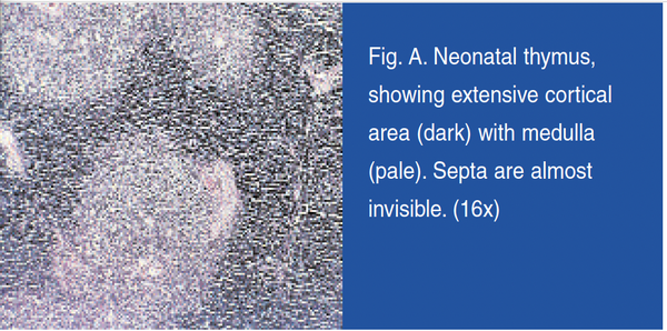
consequence of aging. The following information summarizes the highlights. Thymus Gland Newborn animals possess very active thymus glands, as they function to program a vast number of T lymphocytes and produce high amounts of thymic factors and hormones. Comparison of photomicrographs of neonatal thymus glands (Figure A) and adult bovine thymus (Figure B) dramatically illustrates the differences. The neonatal thymus from newborn
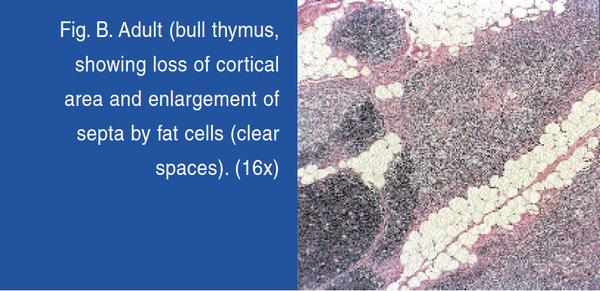
calves possesses abundant cortical tissue, reflecting maximal immune activity. There is very little fat deposition and few Hassall’s capsules, which represent degenerating reticular cells. Cells are compact and possess a high nuclear content, verified by the high level of DNA. In contrast, adult bovine thymus contains massive fatty deposits and there is a minimal cortical area. Most noteworthy is the research that indicates the thymus
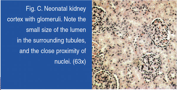
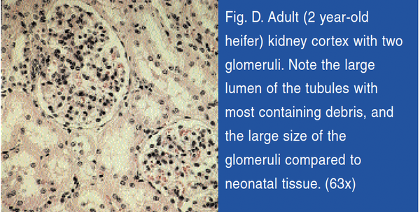
undergoes extensive hypertrophy with aging. Kidneys Small glomeruli with hyperchromic nuclei and small tubules characterize newborn calf kidneys (Figure C). The absence of kidney stones and casts was also observed. In contrast, the adult kidneys possessed a
less compact and more diffuse structure, with occasional casts and kidney stones, indicative of cumulative damage (Figure D). Kidneys of adult animals have processed thousands of gallons of
urine containing a wide
assortment of wastes, including pesticides, industrial pollutants, plant and microbial toxins, drugs, and synthetic feed additives.
Aging dramatically affects kidney function. With aging, a decline in glomerular filtration rate is observed, increasing the susceptibility to renal problems. A decline in renal tubular function has also been documented. Studies of transforming growth factor beta (TGF beta), which limits tissue injury, revealed increased synthesis of TGFs in kidneys of neonatal animals.
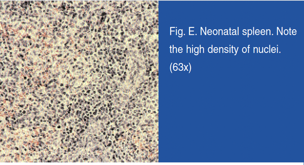
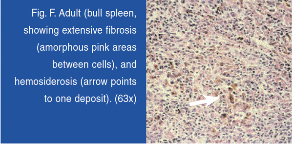
Spleen The neonatal spleen (Figure E) is active in producing and programming lymphocytes, while the adult spleen is less involved. The spleen from newborn calves revealed large lymphoid follicles (Malphigian corpuscles), with a greater concentration of nuclei per cell. Spleen immune function is known to decline with age and with fibrosis. Adult spleens contain smaller lymphoid follicles, and frequently exhibit fibrotic streaks, suggestive of tissue degeneration (Figure F). The neonatal
tissue does not contain brown deposits of hemosiderin, which accumulates in mature spleens. Aging is associated with altered antioxidant defenses in the spleen. Cytoplasmic superoxide
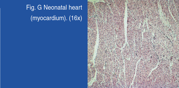
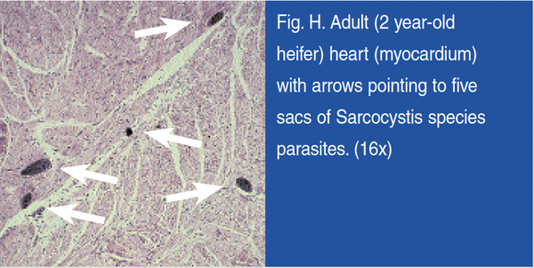
dismutase activity in the spleen of aged rats decreased significantly, when compared to young rats. Age-related decline in cytotoxic lymphocyte activity in mice is associated with changes in both CD8+ and CD4+ cells. Oxidative damage to the spleen is associated with aging. Additionally, age-related loss of immune function has been correlated with the accumulation of oxidative damage in spleen lymphocytes. Heart Histological examination of calf heart did not detect any vascular abnormalities (Figure G). On the other hand, examination of adult bovine heart frequently detected calcification of arteries, and the presence of
numerous parasites (Figure H), confirmation that aging (arteriosclerosis) and
environmental exposure (parasites) are readily detectable in adult bovine hearts. With aging there is a loss of circulatory function, as measured by a drop in V02 max, ranging up to 22% per decade in humans. Age-related decline in function of the heart may be related to increased rates of free radical
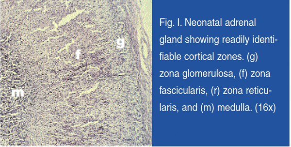
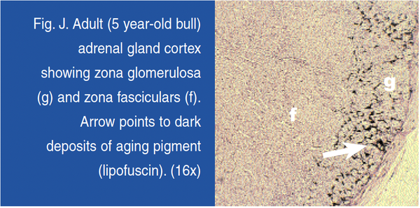
production. Heart mitochondria are the likely subcellular target for increases in susceptibility to free radical attack. Adrenal Glands The adrenal glands of newborn animals revealed distinct, well-defined zones for the cortex and medulla (Figure I), while the adult glands lacked clear functional boundaries and were more diffuse (Figure J).
In addition, the adult cortex exhibited large amounts of lipofuscin, which was absent from neonatal adrenal glands. Lipofuscin
deposits are indicators of lifelong exposure to oxidative stress and lipid peroxidation. Adrenal atrophy and medullary hyperplasia were noted in senescence-accelerated animals, which is evidence of declining adrenal function with advancing age. In
humans there is a marked decline in circulating adrenal 19 carbon steroids and their androgen metabolites with aging, including DHEA and DHEA-sulfate. Possibly the age-dependent decline in glucocorticoid secretion involves impaired conversion of cholesterol to pregnenolone. Additionally, the decline in adrenal cortisol secretion in guinea pigs was demonstrated to correlate closely with decreased adrenal steroid mitochondrial metabolism. Additionally, calf pancreatic enzymes, such as chymotrypsin, elastase, carboxypeptidases and amylase, from milk-fed calves increased in activity after birth until weaning. The secretory activity of the pancreatic islets
has also shown a decline in function with increasing age, as evidenced by fibrosis around the pancreatic islets in aged animals, as compared to islets of younger animals. Amylase output is also markedly higher in younger
animals, which was shown to decrease to below 20% in older animals. Liver The proliferative activity of adult hepatocytes is generally low; histochemical assessment indicate less than 0.1% of hepatocytes in the S phase of the cell cycle. In contrast, dividing cells are common in the liver of newborn animals. The neonatal liver secretes fetal proteins, such as alpha-1 fetoprotein and fetal growth factors, including fetal somatomedin C. These products do not occur in
significant amounts in adult glands. In addition, aging alters the expression of many liver enzymes and proteins. The level of proteins in liver and brain extracts of animals of different ages revealed a progressive oxidation, increasing with age. In the brain and liver the accumulation of oxidized protein corresponded to a loss of key enzyme activities. In older animals oxidized protein may represent between 30 to
50% of the total cellular protein. Brain and Hypothalamus/Pituitary Axis Abnormalities of the hypothalamus-pituitary-adrenal axis regulation are common with aging. For example, regions of the brain showed major deficits in cell signaling in
older animals. In the brain, accumulation of oxidized protein corresponds to a loss of key enzyme activities. Pancreas The postnatal pancreas responds to hormonal
stimulation, for example as triggered by cholecystokinin. The gastrointestinal peptides motilin, cholecystokinin and secretion have been shown to increase pancreatic secretions in preruminant calves. The neonatal pancreas
shows a much stronger expression cytokeratins, as compared to adults. Lower enzyme production in the adult bovine pancreas is also evidenced by the diminished production of the enzyme rennet, an enzyme that curdles
milk, shown to be significantly lower in adult animals,
as compared to a higher prevalence in newborn pancreas. Pesticide Contamination In Bovine Tissues The meat industry reportedly uses hundreds of feed additives for cattle. Among the environmental stressors on adult animals, pesticide exposure is a major concern. Cattle are inevitably exposed to pesticide residues as evidenced by the significant levels of DDT, BHC and dieldrin detected in imported beef. As the liver is the major detoxification site
for many xenobiotics, tissue examination of the liver is a good indicator of pesticide deposition. When analyzed by an independent testing laboratory for approximately forty different chlorinated pesticides and phospho-pesticides, Biotics Research’s neonatal bovine liver powder showed concentrations below the limits of detection for all forty compounds. Other tissues examined in comparison included calf liver and adult beef liver. Contrary to the neonatal
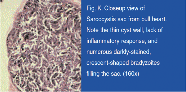
tissue, the adult bovine liver showed detectable amounts of a variety of pesticides. Parasites In Adult Bovine Tissues Pathogenic microorganisms represent another class of environmental stressors to which livestock are exposed. As an example, intestinal sarcocytosis can be a significant health problem if meat is undercooked. Cattle are intermediate hosts
for three species of Sarcocystis cruzi. In one study, bovine tissues from carcasses condemned for eosinophilic myostitis were compared with unaffected animals approved for human consumption. All affected carcasses exhibited granulotomous lesions, and the parasite Sarcocystis cruzi was present in all carcasses. Additionally, carcasses approved for human consumption generally had more infective parasites than condemned carcasses. The parasitic cysts are resistant to both organic solvents and
freeze-drying. Note the readily visible parasites in the adult bovine heart tissue (Figure K). Although the significance of the consumption of adult bovine heart glandulars for human health is unclear, glandular tissues have demonstrated respective organ support. DNA And Protein Content Of Biotics Research Glandular Products DNA is a marker for nuclear material, including histones and nonhistone proteins. When DNA of respective tissues is analyzed, cells from young glands contain a high ratio of nuclear to cytoplasmic volume, as compared to adult glands. DNA analysis, performed by an independent testing laboratory, analyzed the following
glandular preparations: Biotics Research’s Cytozyme-THY™, Cytozyme-AD™ and Cytozyme-SP™, which was compared to Thymus
PMG (protomorphogen), Drenatrophin PMG and Spleen PMG products. The glandular content per tablet varied with the gland and the manufacturer. The results indicated that the DNA content of both manufacturers’ spleen
glandulars was equivalent. The similar DNA levels of Cytozyme-SP™ and Spleen PMG, in all likelihood, reflects
spleen function. That is, as a scavenger of blood cells, the adult spleen (Spleen PMG) might be anticipated to contain high levels of DNA due to its function in mature animals. However, as evidenced in Figures E and F, newborn spleen shows a higher nucleic density, and does not show the extensive fibrosis and hemosiderosis of the adult tissue, which is theoretically a fraction enriched
so-called “protomorphogen”. The “protomorphogen” as an enriched source should contain more DNA than a
preparation of raw whole glands. This clearly is not the case, as the histological data suggests glandulars from
Biotics Research contain twice as much nuclear material (DNA) as the corresponding PMG product, evidenced by the DNA percentage of both Cytozyme-THY™ and Cytozyme-AD™. Conclusion The above synopsis aims to confer the superior nature of Biotics Research Corporation’s neonatal glandulars, by virtue of the histological comparisons. Neonatal glandular therapy, otherwise referred to as organotherapy, may be
incorporated as a comprehensive part of your patient’s nutritional program, as an organ support program, or as an ideal mechanism to support the
respective organ system(s). Obviously, by virtue of the pristine nature of
the neonatal glandulars, they offer a superior adjunct to any nutritional program.
|
May you have great glandular health, Stephen Heuer, B.S. Nutripath
stephen@sygn44.com
|
|
|
|